Bone Cross Section Labeled - Bone Structure Anatomy And Physiology I : This photo shows a cross section through bone.. Shop bone cross section diagram label created by chartsanddiagrams. The central haversian canal, and horizontal canals (perforating/ volkmann's) canals contain blood vessels and nerves from the periosteum. Über 7 millionen englischsprachige bücher. Download 706 bone cross medical section stock illustrations, vectors & clipart for free or amazingly low rates! The wider section at each end of the bone is called the epiphysis (plural = epiphyses), which is filled with spongy bone.
The outlined area is a cross section of an osteon of compact bone. In three dimensions an osteon is cylindrical in shape. Bone in arm pictures 12 photos of the bone in arm pictures bone cancer arm pictures, pictures of bone cancer in arm, bone, bone cancer arm pictures, pictures of bone cancer in arm Bone test anatomy and physiology 12 photos of the bone test anatomy and physiology anatomy and physiology bone lab test, anatomy and physiology bone markings test, anatomy and physiology bone practical test, anatomy and physiology bone tissue test, anatomy and physiology test on bone tissue, bone, anatomy and. Related posts of bone cross section labeled.
5 3 Bone Structure Medicine Libretexts from med.libretexts.org Bones in your body names. They are obtained by taking imaginary slices perpendicular to the main axis of organs, vessels, nerves, bones, soft tissue, or even the entire human body. Bone test anatomy and physiology 12 photos of the bone test anatomy and physiology anatomy and physiology bone lab test, anatomy and physiology bone markings test, anatomy and physiology bone practical test, anatomy and physiology bone tissue test, anatomy and physiology test on bone tissue, bone, anatomy and. This slide contained a cross section of a very small bone, and you are looking at the entire thickness of the shaft of the bone. Bone decalcification is the removal of the mineral component using an acid, leaving the bone soft and easy to cut. 100x first focus in the compact decalcified bone (cb) on the left part of the image, you can see small dots, which are. Bone in arm pictures 12 photos of the bone in arm pictures bone cancer arm pictures, pictures of bone cancer in arm, bone, bone cancer arm pictures, pictures of bone cancer in arm Über 7 millionen englischsprachige bücher.
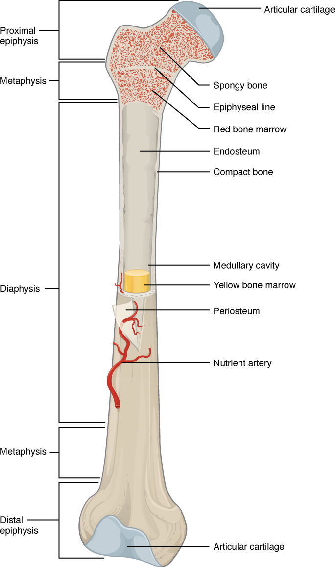
The wider section at each end of the bone is called the epiphysis (plural = epiphyses), which is filled with spongy bone.
The osteocytes are arranged in concentric rings of bone matrix called lamellae (little plates), and their processes run in interconnecting canaliculi. Red bone marrow fills the spaces between the spongy bone in some long bones. Shop bone cross section diagram label created by chartsanddiagrams. They are obtained by taking imaginary slices perpendicular to the main axis of organs, vessels, nerves, bones, soft tissue, or even the entire human body. Smartdraw includes 1000s of professional healthcare and anatomy chart templates that you can modify and make your own. Concentric layers of bone cells (osteocytes) and bone matrix surround the central canal. The wider section at each end of the bone is called the epiphysis (plural = epiphyses), which is filled internally with spongy bone, another type of osseous tissue. The outlined area is a cross section of an osteon of compact bone. Related posts of bone cross section labeled. Red marrow fills the spaces in the spongy bone. This photo shows a cross section through bone. With regards to bone anatomy, some of the parts that can be easily identified include: Slides have to be made this way because the matrix of bone is too hard to be cut with a knife as the other tissues are.
I don't find it enhances the image. Marrow in the shaft of long bones is typically yellow, with red marrow in the head through the cancellous bone. The wider section at each end of the bone is called the epiphysis (plural = epiphyses), which is filled with spongy bone. Compact bone is very different from the other tissues you have seen. This photo shows a cross section through bone.
Bone Structure Anatomy And Physiology I from s3-us-west-2.amazonaws.com Concentric layers of bone cells (osteocytes) and bone matrix surround the central canal. Imaios and selected third parties, use cookies or similar technologies, in particular for audience measurement. The wider section at each end of the bone is called the epiphysis (plural = epiphyses), which is filled internally with spongy bone, another type of osseous tissue. Each epiphysis meets the diaphysis at the metaphysis. Marrow in the shaft of long bones is typically yellow, with red marrow in the head through the cancellous bone. Each of these cylinders is called an osteon or haversian system. It includes such bones as the hip and vertebrae. I don't find it enhances the image.
Bone decalcification is the removal of the mineral component using an acid, leaving the bone soft and easy to cut.
The wider section at each end of the bone is called the epiphysis (plural = epiphyses), which is filled with spongy bone. Related posts of cross section of human bone diagram bone in arm pictures. Each of these cylinders is called an osteon or haversian system. In the center of each osteon is the central canal, a space that houses blood vessels and nerves that supply bone. Fixed slide cross section of muscle tissue, 100x microscope view. Bone in arm pictures 12 photos of the bone in arm pictures bone cancer arm pictures, pictures of bone cancer in arm, bone, bone cancer arm pictures, pictures of bone cancer in arm And why does the marrow stop where it does, and so sharply? Trust pharmacy 221 massachusetts ave, boston, ma 02115. Red marrow fills the spaces in the spongy bone. I don't find it enhances the image. Über 7 millionen englischsprachige bücher. Slides have to be made this way because the matrix of bone is too hard to be cut with a knife as the other tissues are. New users enjoy 60% off.
Would it be a good thing to show the epiphyseal plate? Free online quiz compact bone microscope slide labeled. Bones in your body names. To the left is muscle tissue, and to the right is bone marrow. Imaios and selected third parties, use cookies or similar technologies, in particular for audience measurement.
Bone Structure Anatomy And Physiology I from s3-us-west-2.amazonaws.com They are obtained by taking imaginary slices perpendicular to the main axis of organs, vessels, nerves, bones, soft tissue, or even the entire human body. Browse 4,294 bone cross section stock photos and images available, or search for human bone cross section to find more great stock photos and pictures. At this level of magnification, the fundamental structure of compact bone is visible. Would it be a good thing to show the epiphyseal plate? Bones in your body names. Cross section anatomy of human bone cross section anatomy of human bone. Cookies allow us to analyze and store information such as the characteristics of your device as well as certain personal data (e.g., ip addresses, navigation, usage or geolocation data, unique identifiers). 100x first focus in the compact decalcified bone (cb) on the left part of the image, you can see small dots, which are.
Bone in arm pictures 12 photos of the bone in arm pictures bone cancer arm pictures, pictures of bone cancer in arm, bone, bone cancer arm pictures, pictures of bone cancer in arm
Red marrow fills the spaces in the spongy bone. Red marrow fills the spaces in the spongy bone. Cross section anatomy of human bone cross section anatomy of human bone. Bone decalcification is the removal of the mineral component using an acid, leaving the bone soft and easy to cut. Red bone marrow fills the spaces between the spongy bone in some long bones. Red marrow fills the spaces in the spongy bone. With regards to bone anatomy, some of the parts that can be easily identified include: Related posts of cross section of human bone diagram bone in arm pictures. The wider section at each end of the bone is called the epiphysis (plural = epiphyses), which is filled with spongy bone. Why is the marrow red? At this level of magnification, the fundamental structure of compact bone is visible. Each of these cylinders is called an osteon or haversian system. The wider section at each end of the bone is called the epiphysis (plural = epiphyses), which is filled with spongy bone.
Related posts of bone cross section labeled bone cross section. To the left is muscle tissue, and to the right is bone marrow.


0 Komentar